Saturday, February 16, 2013

Thalasemia


The major hemoglobin in adults is hemoglobin A, which is a tetramer consisting of two pairs of globin polypeptide chains: one pair of alpha chains; and one pair of beta chains. In normal subjects, globin chain synthesis is very tightly controlled so that the ratio of production of alpha to non-alpha chains is 1.00 ± 0.05. There are two copies of the alpha globin gene on chromosome 16. A single beta globin gene resides on chromosome 11 adjacent to genes encoding the beta-like globin chains, delta and gamma.
Thalassemia refers to a spectrum of diseases characterized by reduced or absent production of one or more globin chains. Beta thalassemia is due to impaired production of beta globin chains, which leads to a relative excess of alpha globin chains. These excess alpha globin chains are unstable, incapable of forming soluble tetramers on their own, and precipitate within the cell, leading to a variety of clinical manifestations. The degree of alpha globin chain excess determines the severity of subsequent clinical manifestations, which are profound in patients homozygous for impaired beta globin synthesis and much less pronounced in heterozygotes who generally have minimal or mild anemia and no symptoms. 
Alpha thalassemia, in comparison, is due to impaired production of alpha globin chains, which leads to a relative excess of beta globin chains. The toxicity of the excess beta globin chains in alpha thalassemia on the red cell membrane skeleton appears to be less than that of the excess partially oxidized alpha globin chains in beta thalassemia. This probably explains why the clinical manifestations are generally less severe in alpha compared with beta thalassemia of comparable genetic severity (except for homozygous alpha (0) thalassemia, which is incompatible with extrauterine life, leading to hydrops fetalis and/or death shortly after delivery).


Certain clinical terms are used to describe the phenotypic expression of beta thalassemia:
  • Beta (0) thalassemia — Beta (0) thalassemia refers to mutations of the beta globin locus that result in the absence of production of beta globin. Patients homozygous or doubly heterozygous for beta (0) thalassemic genes cannot make normal beta chains and are therefore unable to make any hemoglobin A.
  • Beta (+) thalassemia — Beta (+) thalassemia refers to mutations that result in decreased production of beta globin. Patients homozygous for beta (+) thalassemic genes are able to make some hemoglobin A, and are generally less severely affected than those homozygous for beta (0) genes.
  • Beta thalassemia major — Beta thalassemia major is the term applied to patients who have either no effective production (as in homozygous beta (0) thalassemia) or severely limited production of beta globin. These are the patients originally described by Cooley (Cooley's anemia). Starting during the first year of life, they have profound and life-long transfusion-dependent anemia.
  • Beta thalassemia minor — Beta thalassemia minor (beta thalassemia trait) is the term applied to heterozygotes who have inherited a single gene leading to reduced beta globin production. Such patients are asymptomatic, may be only mildly anemic, and are usually discovered when a blood count has been obtained for other reasons.
  • Beta thalassemia intermedia — Beta thalassemia intermedia is the term applied to patients with disease of intermediate severity, such as those who are compound heterozygotes of two thalassemic variants (table 1). These patients have a later clinical onset and a milder degree of anemia, which may or may not require transfusional support.
STAGES OF INCREASED ICP


Stages of intracranial hypertension

Minimal increases in ICP due to compensatory mechanisms is known as stage 1 of intracranial hypertension.

When the lesion volume continues to increase beyond the point of compensation, the ICP has no other resource, but to increase. Any change in volume greater than 100–120 mL would mean a drastic increase in ICP. This is stage 2 of intracranial hypertension.
Characteristics of stage 2 of intracranial hypertension include compromise of neuronal oxygenation and systemic arteriolar vasoconstriction to increase MAP and CPP.

Stage 3 intracranial hypertension is characterised by a sustained increased ICP, with dramatic changes in ICP with small changes in volume.
In stage 3, as the ICP approaches the MAP, it becomes more and more difficult to squeeze blood into the intracranial space. The body’s response to a decrease in CPP is to raise blood pressure and dilate blood vessels in the brain. This results in increased cerebral blood volume, which increases ICP, lowering CPP and perpetuating this vicious cycle.
This results in widespread reduction in cerebral flow and perfusion, eventually leading to ischemia and brain infarction.

Neurologic changes seen in increased ICP are mostly due to hypoxia and hypercapnea and are as follows:
1. Decreased level of consciousness (LOC),
2. Cheyne–Stokes respiration,
3. Hyperventilation,
4. Sluggish dilated pupils and
5. Widened pulse pressure.

Monday, February 11, 2013

Blood Transfusion


Transfusion Technique:
- Maximum time over which blood products can be administered is 4 hrs for 1 unit because of danger of bacterial proliferation & RBC hemolysis;

- If slower infusion rate is required, half of the unit may be infused while other portion remains refrigerated in the blood bank;

- If flow rate is interrupted for >30 minutes, unit must be discarded;

- Blood should be administered through 170 um filters to prevent infusion of macro-aggregates of fibrin and debris as well as leukocytes; 

- Patients should be observed for the first 5-10 min of a transfusion and then examined frequently for signs of fluid overload and other adverse reactions;

- Emergent transfusion:

- In most cases an Rh type and screen takes 10 minutes and is safer than using O negative blood;


Characteristics of pRBC;
    - approx 300 +/- 25 mL
   
 - hematocrit: 70 +/- 5%;

 - one unit of pRBCs should increase hemoglobin by approximately 1 gm/dl;

 - citrate is used as an anticoagulant in blood products during plasmapheresis;
    
- citrate is converted to bicarb by liver & causes metabolic alkalosis

- induction of a metabolic alkalosis may produce an abrupt increase in the  hemoglobinn oxygen affinity;


- w/ transfusion actual amount of potassium administered is approx between 5.2 to  6.6 mEq per unit of pRBC;


- since the mean age of blood administered to trauma pts is 13.5 days (and not 35 to  49 days - expiration date of blood), the actual amount of potassium administered per  unit may be only 1 to 3 mEq;


- w/ massive transfusion hypokalemia is more frequently encountered than  hyperkalemia;- this may be also due to alkalosis (from citrate)


DPG:

- w/ blood that is stored in acid citrate dextrose (ACD) solution for upto to three  weeks is based on the survival of at least 70% of cells in recipients circulation;

- during 3 week period, there is decline in  2-3 disphosphoglycerate (DPG) and a  progressive increase in hemoglobin oxygen affinity (left shift of the oxygen   dissociation curve);


- after transfusion DPG levels require 24 hours or longer to return nl;


- Citrate toxicity:

- can be prevented or its effects minimized by the administration of Ca;

- historically 1gm of CaCl has been given for every four units of blood administered until such time as the pt is normothermic, euvolemic, and is known to have reasonably normal hepatic function;

- if Ca gluconate is used, dose must be 4 times greater than w/ CaCl;

- improved approach is to measure the ionized calcium level.


Complications of Blood Transfusion:
- Blood Product Menu:
- pRBCs - Fresh Frozen Plasma - Platlets - Cryoprecipitate - Transfusion Therapy - Coag Pathway
- Acidemia and Hyperkalemia: from massive transfusions;
- massive transfusion: transfusion of pRBC >6-8 units, must also provide platlets;
            - 8 units platlets for ea 10-12 units pRBC's transfused;
            - 2 units of FFP
            - Ca replacement if hypocalcemic (2nd to citrate)
            - references:
                   
- Electrolyte and acid-base disturbances caused by blood transfusions.
- Hyperkalemia after packed red blood cell transfusion in trauma patients.
Post-Transfusion Alkalosis:
           - the early net result of successful resuscitation is post-transfusion alkalosis in 
            the patient;
           - the sodium citrate is converted to bicarbonate
           - the alkalosis is associated with increased potassium excretion;
Hypocalcemia:
           - some recommend calcium supplementation for patients receiving greater 
            than 100 ml/min;
           - give 0.2 gm of CaCl in a separate line for each 500 ml given;
           - some believe that most patients will tolerate 1 unit pRBC q 5 min without 
             requiring calcium supplementation;
Hemolysis:
    - Non hemolytic reaction:     
           - typically, this reaction occurs after a significant portion of the blood has 
            already been transfused;
           - note: hives + hypotension = anaphylaxis
           - management:
                  - by itself, may continue the transfusion (benadryl 50mg PO/IV);
                  - prior to future transfusions, the patient should be pre-medicated 
                   w/ benadryl 50mg PO/IV (not IM);
                  - if this fails to prevent urticarial rxn, washed RBC's should be
                   given;
                  - w/ mild febrile transfusion reactions fever w/o evidence of 
                   hemolysis or more severe symptoms), antipyretics can be used;
    - Acute hemolytic reaction:
           - most severe and potentially dangerous transfusion reactions;
           - acute intravascular hemolysis occurs during or shortly after transfusion of 
            incompatible blood and is usually due to preformed antibodies; 
                  - typically this reaction occurs early w/ as little as 30 cc of 
                   transfused blood;
           - Manifestations:  
                  - fever, chills, back or chest pain, N/V, and evidence of 
                    hemodynamic instability;
           - Required labs:
                  - spin a hematocrit to look for a pink plasma layer indicates 
                    hemolysis;
                  - pink-red (spun) plasma indicates that greater than 20 mg/dl of 
                   free hemoglobin is present;
                  - send off a DIC screen: PT/PTT, fibrinogen, fibrinogen
                   degradation products, serum bilirubin;
                  - culture of the patient and the donor blood is indicated if there is 
                    suspicion of bacterial contamination;
                  - repeat cross match;
                  - Coomb's Test, Free Hb;
                  - CBC, RBC morphology;
                  - send Donor's Blood back to the blood back;
                  - repeat cross match;
           - Management:
                  - try to preserve intravascular volume and protect against acute
                   renal failure;
                         - NS 500 ml IV "wide open"
                  - monitor the urine output closely and maintain a brisk diuresis 
                   (greater than 100 ml/hr);
                  - consider alkalinization of the urine with bicarbonate (1 mEq/kg 
                   IV until urine pH =7.5-9.0)
                         - will facilitate the excretion of free hemoglobin 
           - Reference:
                  - Extracorporeal hemolysis in orthopedic patients. Report of two 
                   cases.
- Transmission of disease:
- Increased infection rate:
- septic reaction: considered when high fever and hypotension accompany a 
 transfusion reaction;

Friday, February 8, 2013

Immunostimulant for treatment of Malignant melanoma ?


A) Levamisol
B) BCG
C) Aldesleukin
D) Methotrexate


Ans: ?C (Aldesleukin)
Used as an adjunct in metastatic renal cell carcinoma, recently approved for Tx of metastatic melanoma.

Aldesleukin toxicity:

Treatment associated w/ serious cardiovascular toxicity resulting from capillary leak syndrome
involves loss of vascular tone, leakage of plasma proteins, adn fluid into extravascular space
may result in hypotension, reduced organ perfusion and even death.


A. Levamisole: 
Antiparasitic drug that stimulates maturation/ proliferation of T cells, enhances T- cell mediated immune responses.
Adjunctive treatment together w/ 5-FU and leucovorin after surgical resection in patients w/ dukes stage C colon cancer.
Has been used in treatment of nephrotic syndome.
Adverse effects: neutorpenia, anemia, thrombocytopenia, agranulocytosis, encephalopathy assoc. w/ demyelination.


B. BCG: 
Bacillus-calmette-guerin used as a non specific immunostimulant.
Used for intravesical therapy of bladder cancer.


D. Methotrexate 
It is an antimetabolite and antifolate drug. It is used in treatment of cancer, autoimmune diseases, ectopic pregnancy, and for the induction of medical abortions. It acts by inhibiting the metabolism of folic acid.


Interferons beta 1a
Iinterferon beta 1b
Approved for use in MS


Interferons gamma B

Used in chronic granulomatous disease, activates phagocytes.


Oprelvekin:

Recombinant form of human IL 11 derived from genetically altered E. coli, stimulates platelet formation.
Used to treat thrombocytopenia.


Filgrastim:

Granulocyte colony stimulating factor- a 175 aminoacid glycoprotein produced by e. coli, stimulates CFU-G to increase neutrophil prodution. It is a recombinant, many valency, polypeptide. Lineage-specific hematopoietic agent, works on one cell line, stimulates peripehral blood stem cells.
Used to stimulate bone marrow recovery during cancer chemotherapy.


Sargramostim:

Derived from yeast, nonlineage specific hematopoetic agent as it stimulates both granulocytic and macrophage progenitor cells and the mature cells. Receptors on these cells to which sargramostim binds
similar to endogenous cytokine GM-CSF differing only in one amino acid.
Promotes myeloid recovery in patients given high dose chemotherapy for:

Non-hodgkins lymphoma
Acute lymphoblastic leukemia
Hodgkins disease patients who are undergoing bone marrow transplantation
Used to promote myeloid recovery after standard dose chemotherapy
Used to help myeloid recovery after BMT
To treat neutropenia associated w/ AIDS.

Dose related effects:

May be difficult to separate effects due to endogenous GM-CSF and exogenous Sargramostim.
High doses assoc. w/ bone pain, flu-like symptoms, fever, diarrhea, N and V, rash.
* contraindicated in patients w/ heart failure, pulmonary edema, and yeast hypersensitivity.


Epoetin- alpha (erythropoetin):
Recombinant human growth factor responsible for stimulating comitted erythroid precursors resulting in erythropoesis.

Recominent erythropoietin therapy in conjunction w/ adequate iron intake is used in the treatment w/ anemia assoc w/ :
Surgery
Treatment w/ zidovudine-induced anemia in AIDs patients
Cancer chemotherapy (myelosuppressive agents, when chemotherapeutic agents are highly nephrotoxic they may cause anemia)
Prematurity
Treatment w/ anemia assoc. w/ chronic renal disease including renal failure.


Thrombopoetin:

Myeloid cytokine growth factor is distinct from but analogous to erythropoietin, G-CSF and GM-CSF
cloning and expression of recombinant human thrombopoietin as a fusion protein combining GM-CSF and IL 3 has been very important for patients whose boen marrow has been compromised by chemo
selectively stimulates megakaryocytopoiesis.

Recombinant fusion protein-thrombopoetin + GM-CSF + IL 3 is used to treat anemia, neutropenia, and thrombocytopenia assoc. w/ high dose chemo.
Recombinant human thrombopoetin is used in accelerating platelet recovery in patients undergoing hematopoetic stem cell transplantation.


Thursday, February 7, 2013

Derma Notes


The lower epidermal cells (basal layer) produce a variety of keratin filaments and desmosomal proteins (e.g.
desmoglein and desmoplakin), which make up the ‘cytoskeleton’.
This confers strength to the epidermis and prevents it shedding off.

Higher up in the granular layer, complex lipids are secreted by the keratinocytes and these form into intercellular lipid bilayers, which act as a semipermeable skin barrier.

The upper cells (stratum corneum) lose their nuclei and become surrounded by a tough impermeable ‘envelope’


GLP-1 IN T2DM


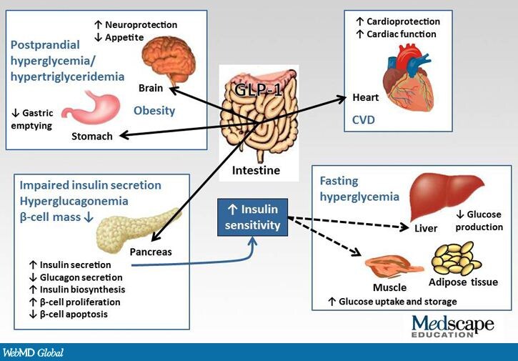
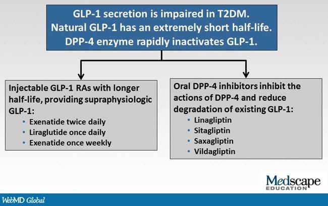
Several antihyperglycemic agents have been developed in order to address some of the challenges associated with older agents. Incretin-based therapies emerged in the past decade and have become established as important treatment options for patients with T2DM. Two classes of agents are available: (1) DPP-4 inhibitors, which are administered orally and include sitagliptin, saxagliptin, vildagliptin, and linagliptin; and (2) GLP-1 RAs, which are administered subcutaneously and include exenatide twice-daily, exenatide once-weekly, and liraglutide.

Sodium glucose cotransporter 1 and 2 inhibitors are another class of novel antihyperglycemic agents. They inhibit glucose reabsorption from renal tubules and cause glucosuria, and sodium glucose cotransporter 1 inhibitors also lead to reduced uptake of glucose from the small intestine. Numerous sodium glucose cotransporter 2 inhibitors are undergoing preclinical and clinical trials, with dapagliflozin having completed phase 3 trials. It has been shown to reduce A1c, reduce blood glucose levels, and is associated with weight loss.


Saturday, February 2, 2013

RA TREATMENT IN PREGNANCY

Methotrexate should be avoided in women trying to conceive or who are pregnant since it is a proven teratogen (pregnancy category X). The data on biologics during pregnancy is much less clear. The anti-TNF therapies are all pregnancy category B. The other commonly used biologics in RA (abatacept, rituximab, tocilizumab) are pregnancy category C. The Food and Drug Administration (FDA) currently warns against using biologics in pregnancy. For women, who require therapy during pregnancy, glucocorticoids, hydroxychloroquine, and occasionally azathioprine (after the first trimester) are the preferred therapies for most physicians who have experience prescribing these agents during pregnancy.


Methotrexate is to be avoided, in particular, since it is a proven teratogen (pregnancy category X). It should be stopped for at least 1 menstrual cycle and as much as 6 months prior to attempting to conceive. Men with RA on methotrexate should also discontinue methotrexate for 3 months or more prior to conception. The data on biologics during pregnancy are much less clear. There have been case series describing various congenital anomalies affecting fetuses of women using these agents, and there have also been anecdotal reports of women experiencing normal pregnancies and good fetal outcomes with exposure to these compounds.[20] The anti-TNF therapies are all pregnancy category B (animal reproduction studies have failed to demonstrate a risk to the fetus, and there are no adequate and well-controlled studies in pregnant women; or animal studies have shown an adverse effect, but adequate and well-controlled studies in pregnant women have failed to demonstrate a risk to the fetus in any trimester). The other commonly used biologics for RA (abatacept, rituximab, tocilizumab) are pregnancy category C (animal reproduction studies have shown an adverse effect on the fetus and there are no adequate and well-controlled studies in humans, but potential benefits may warrant use of the drug in pregnant women despite potential risks).


Instruments to Measure Rheumatoid Arthritis Disease Activity and Define Remission


Patient Activity Scale (PAS) or PAS-II (range: 0-10)
Remission: 0 to 0.25
Low activity: 0.26 to 3.7
Moderate activity: 3.71 to <8.0
High activity: ≥8.0


Routine Assessment of Patient Index Data 3 (range: 0-10)
Remission: 0 to 1.0
Low activity: >1.0 to 2.0
Moderate activity: >2.0 to 4.0
High activity: >4.0 to 10


Clinical Disease Activity Index (range: 0 -76.0)
Remission: ≤2.8
Low activity: >2.8 to 10.0
Moderate activity: >10.0 to 22.0
High activity: >22


Disease Activity Score in 28 joints (range: 0- 9.4)
Remission: <2.6
Low activity: ≥2.6 to <3.2
Moderate activity: ≥3.2 to ≤5.1
High activity: >5.1


Simplified Disease Activity Index (range: 0- 86.0)
Remission: ≤3.3
Low activity: >3.3 to ≤11.0
Moderate activity: >11.0 to ≤26
High activity: >26


Rheumatoid arthritis (RA)

Rheumatoid arthritis (RA) has a typical onset in middle age with a stronger predominance in women. Testing for serum RF and anticyclic citrullinated peptide (CCP) antibodies, a test with higher specificity, may help corroborate or provide a harbinger for clinical diagnosis of RA. It should be noted that serum RF may not be positive in the early stages of RA, in contrast to the anti-CCP antibody, which may predate full-blown RA by months to years.

In regards to the differential diagnosis of RA, various forms of viral arthritis, most notably, arthritis due to parvovirus B19, hepatitis B (in the anicteric phase), hepatitis C, and human immunodeficiency virus (HIV), can present with sudden onset joint pain and/or swelling.

Early disease modifying antirheumatic drug (DMARD) therapy in RA substantially improves signs and symptoms of the disease, limits joint damage, and improves outcomes and long-term prognosis. Methotrexate is the most common first DMARD initiated for RA, and a typical starting dose is between 7.5 to 15 mg weekly. Methotrexate is optimally administered with daily folic acid supplementation. While nonsteroidal anti-inflammatory drugs (NSAIDs), like diclofenac, may provide relief of signs and symptoms, they do not improve outcomes or prognosis in RA.


Disease modifying antirheumatic drug therapy should be started early, ideally within 3 months from the onset of symptoms. The most commonly initiated first DMARD for RA is methotrexate. With the initiation of methotrexate, patients should have baseline liver enzymes and a complete blood count (CBC), and these tests should be monitored at regular intervals to exclude hematologic or hepatic toxicity attributable to methotrexate.


The 2012 ACR recommendations suggest to measure RA disease activity in a quantitative way, and to therapeutically aim for a target of remission or low disease activity; however the recommendations do not specify which specific measurement tools to use.
The RAPID3 (Routine Assessment of Patient Index Data 3 ),
CDAI (Clinical Disease Activity Index),
DAS28 (Disease Activity Score in 28 joints with ESR), and
SDAI (Simplified Disease Activity Index)
are all mentioned in the ACR guidelines, however use of any of these measures as well as the HAQ-DI (Health Assessment Questionnaire Disability Index) are appropriate to use as long as they are used consistently to follow RA disease activity over time.


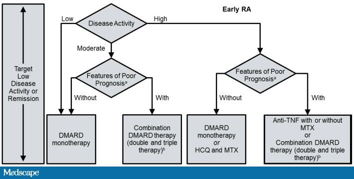
2012 ACR Recommendations for the Treatment of Early RA (<6 Months)


ACR = American College of Rheumatology; CCP = cyclic citrullinated peptide; DMARD = disease modifying antirheumatic drug; HAQ-DI = Health Assessment Questionnaire-Disability Index; HCQ = hydroxychloroquine; LEF = leflunomide; MTX = methotrexate; RA = rheumatoid arthritis; TNF = tumor necrosis factor.
aPatients were categorized based on the presence or absence of 1 or more of the following poor prognostic features: functional limitation (eg, HAQ-DI or similar validated tools), extra-articular disease (eg, presence of rheumatoid nodules, RA vasculitis, Felty’s syndrome), positive rheumatoid factor or anti-CCP antibodies, and bony erosions by radiograph.
bCombination DMARD therapy with 2 DMARDs, which is most commonly MTX-based with some exceptions (eg, MTX+HCQ, MTX+LEF, MTX+sulfasalazine, and sulfasalazine+HCQ), and triple therapy (MTX+HCQ+sulfasalazine).


The majority of the clinical trials with the anti-TNF agents have examined patients who had previously failed treatment with nonbiologic DMARDs, where biologic agents were added to methotrexate. These studies have consistently demonstrated, largely across the different anti-TNF agents, that anti-TNF agents have superior efficacy in terms of disease activity and retarding progression of radiographic damage (bone erosions and joint space narrowing) compared to the use of methotrexate alone.


Anti-TNF agents have been associated with a heightened risk for both typical and atypical infections based on both clinical trials and large observational studies. The 2012 ACR guidelines recommend the use of either a TB skin test (purified protein derivative [PPD]) or an interferon gamma release assay (IGRA), commonly called a quantiferon test, for TB screening prior to use of biologic agents.
The IGRA has a particular advantage in patients who have been vaccinated in the past with the Bacillus Calmette-Guérin vaccine.


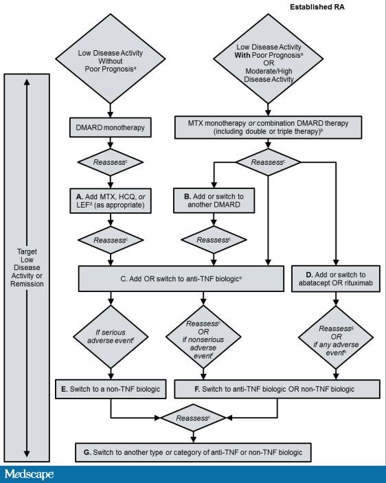
Evidence has not shown increased efficacy for doubling the frequency of dosing for anti-TNF therapy (i.e if patient having moderate disease activity even with alternate weekly dose of adalimumab 40) and the combination of biologics has been associated with unacceptable toxicity. According to the guidelines, this patient should be switched to either a different anti-TNF agent or to a non-TNF biologic agent (abatacept, rituximab, or tocilizumab)


2012 ACR Recommendations for the Treatment of Established RA Disease (Duration ≥6 Months).


There have been several studies that have examined the additive use of biologic agents and while there may be some synergistic benefit in terms of efficacy, the toxicity, particularly from infections, has been considered unacceptable for the use of combinations of biologic agents. Thus, combination biologic therapy (eg, abatacept plus an anti-TNF agent ) is not recommended based upon the available evidence.


The 2012 ACR guidelines recommend against administering live virus vaccinations including influenza vaccine administered by nasal spray, herpes zoster vaccination, and yellow fever vaccine in patients currently on biologic therapy. Inactivated vaccines including injectable influenza and pneumococcal vaccinations may safely be given to patients receiving biologic agents and nonbiologic DMARDs.

2012 ACR recommendations Regarding the Use of Vaccines in Patients With RA Starting or Currently Receiving DMARDs or Biologic Agents

 Killed VaccinesRecombinant VaccineLive Attenuated Vaccine
 PneumococcalaInfluenza (intramuscular)Hepatitis BbHuman papillomavirusHerpes zoster
Before initiating therapy:     
    DMARD
    monotherapy
    Combination
    DMARDs
    Anti-TNF
    biologics
    Non-TNF
    biologics
While already taking therapy:     
    DMARD
    monotherapy
    Combination
    DMARDs
    Anti-TNF
    biologics
Not recommended
    Non-TNF
    biologics
Not recommended


Post-meal time glucose spikes are to be prevented because:


  • Lowering A1c reduces microvascular complications
  • A1c reflects PPG as well as fasting plasma glucose levels (individuals are “postprandial” most of the 24-hour period)
  • Control of PPG levels reduces intima-media thickness and decreases cardiac events (as shown in the STOP-NIDDM and other trials)
  • Elevated PPG levels are a highly prevalent issue, especially in the elderly.
  • Abundant experimental evidence relates acute hyperglycemia and glucose excursions to endothelial dysfunction and increased oxidative load.
  • Elevated blood glucose levels 2 hours after a glucose load are associated with an increased risk for death, independent of fasting blood glucose levels (even in non-diabetic persons). The European Prospective Investigation into Cancer Study–Norfolk cohort study further raises questions as to the level of A1c associated with increased risk.

Estimated Average Glucose


Table. Estimated Average Glucose
A1c (%)mg/dL* (95% confidence intervals)mmol/L
(95% confidence intervals)
6126 (100-152)7.0 (5.5-8.5)
7154 (123-185)8.6 (6.8-10.3)
8183 (147-217)10.2 (8.1-12.1)
9212 (170-249)11.8 (9.4-13.9)
10240 (193-282)13.4 (10.7-15.7)
11269 (217-314)14.9 (12.0-17.5)
12298 (240-347)16.5 (13.3-19.3)

Principles of the AACE/ACE Algorithm for Glycemic Control


Principles of the AACE/ACE Algorithm for Glycemic Control


  • Early aggressive glucose control, the importance of which has been demonstrated by the DCCT/EDIC trial
  • Targeting PPG excursions, particularly at lower A1c levels (where they comprise a significant component of the A1c)
  • Stratified treatment protocols based on presenting A1c in drug naïve patients or A1c on treatment when not at goal
  • Combination therapy early in the treatment paradigm initially at an A1c of 7.6%-9.0% or after 2 to 3 months of monotherapy at an A1c of 6.5%-7.5%
  • Advancing therapy at 2- to 3-month intervals, addressing the problem of clinical inertia


ADA practice recommendations regarding the prevention and management of diabetes complications


In light of the above strong evidence linking diabetes and CVD and to control and prevent the microvascular complications of diabetes, the ADA has issued practice recommendations regarding the prevention and management of diabetes complications.

Blood pressure should be measured routinely. Goal blood pressure is < 130/80 mmHg. Patients with a blood pressure ≥ 140/90 mmHg should be treated with drug therapy in addition to diet and lifestyle modification. Patients with a blood pressure of 130-139/80-89 mmHg may attempt a trial of lifestyle and behavioral therapy for 3 months and then receive pharmacological therapy if their goal blood pressure is not achieved. Initial drug therapy should be with a drug shown to decrease CVD risk, but all patients with diabetes and hypertension should receive an ACE inhibitor or ARB in their antihypertensive regimen.

Lipid testing should be performed in patients with diabetes at least annually. Lipid goals for adults with diabetes should be an LDL < 100 mg/dl (or < 70 mg/dl in patients with overt CVD), HDL > 50 mg/dl, and fasting triglycerides < 150 mg/dl. All patients with diabetes should be encouraged to limit consumption of saturated fat, trans fat, and cholesterol. Statin therapy to lower LDL by 30-40% regardless of baseline is recommended to decrease the risk of CVD in patients > 40 years of age. Patients < 40 years of age may also be considered for therapy. In individuals with overt CVD, special attention should be paid to treatment to lower triglycerides or raise HDL. Combination therapy with a statin plus other drugs, such as fibrates or niacin, may be necessary to achieve ideal lipid control, but patients should be monitored closely for possible adverse reactions of therapy.

Aspirin therapy (75-162 mg/day) is indicated in secondary prevention of CVD and should be used in patients with diabetes who are > 40 years of age and in those who are 30-40 years of age if other risk factors are present. Patients < 21 years of age should not receive aspirin therapy because of the risk of Reye's syndrome. Patients who cannot tolerate aspirin therapy because of allergy or adverse reaction may be considered for other antiplatelet agents.

In addition to the above pharmacological recommendations, patients with diabetes should be encouraged to not begin smoking or to stop smoking to decrease their risk of CVD and benefit their health in other ways. It should also be noted that statins, ACE inhibitors, and ARBs are strongly contraindicated in pregnancy.

Micro & Macrovascular Diseases

Microvascular disease is a process through which the very small branches of arteries throughout the body become damaged.  Microvascular disease is a common component of other conditions, such as diabetes mellitus and autoimmune diseases.

The very small branches of the arteries are delicate but very important structures.  Damage to these vessels results in occlusion of the vessels and impairment of blood flow.  In many situations the small arteries can re-grow and overcome the blockage, a process called angiogenesis.  This is part of the normal healing process.  In microvascular disease the commonest cause is chemicals within the blood that damage the very delicate lining of the small arteries and causes the blood to clot in the artery and block it.  Sometimes these chemicals are produced by the body itself as part of the immune response and is called an autoimmune microvascular disease.  Occasionally microvascular disease is the result of abnormalities in the cells that form part of the blood.

The commonest symptoms are pain and discoloration of the extremities, usually the fingers and toes, sometimes even leading to gangrene.  These symptoms are very similar to those cause by occlusion of the larger arteries except that it is not associated with muscle pain on exercise (intermittent claudication) and the blood pressure in the larger arteries is normal.

Microvascular disease usually affects the whole body to some degree and the most serious complications are caused by damage to the vital organs (e.g. heart, brain, kidneys, liver).
Treatment for microvascular disease is directed at the underlying cause.  Lifestyle changes to eliminate factors that aggravate the condition, such as smoking, should be the first line of treatment.  A complete medical assessment is required to identify the underlying cause if possible.  If there is an autoimmune element to the condition, then referral to a rheumatologist may be required.  If there is an abnormality in the blood referral to a haematologist is required. Surgery plays only a secondary role in the management of microvascular disease.

  • Diabetic retinopathy
  • Diabetic nephropathy
  • Diabetic neuropathy


Macrovascular disease is a disease of any large (macro) blood vessels in the body. It is a disease of the large blood vessels, including the coronary arteries, the aorta, and the sizable arteries in the brain and in the limbs.
This sometimes occurs when a person has had diabetes for a long time. Fat and blood clots build up in the large blood vessels and stick to the vessel walls.
Three common macrovascular diseases are coronary disease (in the heart), cerebrovascular disease (in the brain), and peripheral vascular disease (in the limbs)

Diabetic treatment

If your A1c is under 7.5%, you could technically use 1 drug. If it is a post-meal problem, use an incretin mimetic or something similar. If the A1c is just under 7.5% and there is a high fasting, use 1 drug to treat that such as metformin.

The ADA suggests maintaining FBG values between 70 and 130 mg/dL and peak post-prandial glucose (PPG) -- 1 to 2 hours following the meal -- values below 180 mg/dL in order to achieve an A1c goal of less than 7%.
Alternatively, the American Association of Clinical Endocrinologists (AACE) recommends a lower A1c target of 6.5%, by maintaining FBG below 110 mg/dL and 2-hour PPG levels below 140 mg/dL. Target A1c levels need to be determined on the basis on several factors, including hypoglycemia risk and vulnerability.


The usual approach in insulin initiation has been the “Fix Fasting First” approach; this is achieved through the use of appropriate basal insulin replacement (usually with basal insulin doses ranging between 0.4 and 0.6 units/kg/day) and the subsequent addition -- if A1c levels remain above target -- of 1 or more doses of a rapid-acting insulin analog added to 1 or more meals in a sequential manner.
Specifically, 4 to 6 units of a rapid-acting analog are added before the main (largest) meal of the day and titrated to either 2-hour PPG (blood glucose < 180 mg/dL) or next preprandial (blood glucose < 130 mg/dL) targets. On average, 8 to 12 units of rapid-acting insulin are needed to adequately control PPG in the average 100-kg patient with type 2 diabetes. If full basal/bolus insulin therapy is needed in patients with type 2 diabetes, then fixed doses of premeal insulin appear to have similar effectiveness and safety as compared to more complex approaches that require matching insulin to carbohydrates or using frequent supplemental scale corrections.

An alternative approach to controlling prandial glycemia, while potentially minimizing both weight gain and hypoglycemia risk, is the use of incretin-based drugs[36] either alone or in combination with basal insulin. Because these pharmacotherapies exert their effect on β-cells (stimulating insulin secretion and release or suppressing glucagon release) in a glucose-dependent manner, they are associated with a low risk for hypoglycemia when used without hypoglycemic agents (eg, sulfonylureas, glinides, insulin).

The 2 major classes of incretin drugs available for clinical use in the United States are dipeptidyl peptidase (DPP)-4 inhibitors (ie, sitagliptin, saxagliptin, linagliptin) and glucagon-like peptide (GLP)-1 receptor agonists (ie, exenatide, liraglutide).
On average, DPP-4 inhibitors lower A1c levels less -- decrease of 0.5% to 0.8% -- than GLP-1 receptor agonists -- decrease of 0.6% to 1.9%. GLP-1 receptor agonists can be further subdivided into short-acting (exenatide) and long-acting (liraglutide long-acting release and exenatide long-acting release) agonists.
On average, short-acting GLP-1 receptor agonists tend to lower PPG more effectively than long-acting GLP-1 receptor agonists, while long-acting GLP-1 receptor agonists have a greater impact on fasting plasma glucose.


PPG levels apparently have a better correlation with A1c than do fasting blood glucose (FBG) levels, especially when the A1c is less than 8.4%. Furthermore, reduced early and “first-phase” insulin releases, fundamental defects in type 2 diabetes, lead to high PPG levels. Insulin resistance and the reduced incretin effect in type 2 diabetes also contribute significantly to postprandial hyperglycemia.


The DCCT/EDIC trial reported a macrovascular benefit in the original intensively controlled group despite subsequent loss of A1c control equivalent to the post DCCT conventionally treated group. This important concept, termed “metabolic memory,” provides evidence supporting the importance of early, aggressive control of diabetes. Additionally, evidence from the Kumamoto Study and the United Kingdom Prospective Diabetes Study confirm reduced microvascular complications with lower A1c levels.


Strong epidemiologic evidence from varied populations links post-challenge hyperglycemia to macrovascular complications; these studies include the Rancho Bernardo Study, the Honolulu Heart Study, the Paris Prospective Heart Study, the Diabetes Intervention Study, and the Diabetes Epidemiology:Collaborative Analysis of Diagnostic Criteria in Europe Study.


Possible effects of acute hyperglycemia responsible for increased microvascular and macrovascular risk include:

  • Endothelial dysfunction
  • Increased oxidative load
  • A pro-inflammatory state
  • Protein glycosylation
  • Altered coagulation

In 2003, Monnier and colleagues demonstrated that, in patients with an A1c level less than 7.3%, reductions in postprandial glycemia were approximately 70% responsible for the improvement in glycemic control as measured by A1c. Whereas for patients with an A1c greater than 8.0%, improvements in fasting glycemic control contributed more to the overall glycemic control than did postprandial glycemia. This observation, practically stated, supports the “Fix Fasting First” approach commonly used in clinical practice.
In 2007, Monnier and colleagues designed a study in early diabetes to demonstrate that the loss of glycemic control progressed sequentially from postprandial to fasting. In this study, the researchers found that the initial differences in mean glucose concentrations reached statistical significance during daytime postprandial periods first, followed by morning periods (otherwise known as the dawn phenomenon), and then nocturnal fasting periods. The finding led Monnier and his team to conclude that the deterioration in glucose control progressed stepwise in the 3 postmeal periods leading to fasting hyperglycemia; this observation, therefore, supports a stepwise treatment program and medication titration.
In the appropriate patients, as determined by their fasting blood glucose (FBG) and A1c, a clinician can effectively titrate treatment to achieve FBG in the target (100 to 126 mg/dL) range. If the A1c remains above the target, then management of the postprandial blood glucose component becomes essential to improve glycemic control.
In a more recent study, Monnier and colleagues suggest that, to achieve glycemic targets less than 6.5% (as currently recommended by ACE/AACE and the International Diabetes Federation), attention must be paid to the PPG. However, there is a caveat to take into consideration: Riddle and colleagues recently published a study evaluating PPG and from their data, it can be concluded that contribution of PPG and fasting plasma glucose depends on treatment.

If the FBG has been optimized or treated to the target and the A1c is still not at goal, targeting the PPG is the next step. A reasonable approach is to start with the largest meal first (typically dinner) and measure the blood glucose 2 hours after the beginning of the meal. Different organizations have proposed different targets.

Friday, February 1, 2013

Influenza treatment

The CDC recommends that antiviral treatment be initiated within 48 hours of the onset of symptoms if possible. Early treatment can shorten the duration of illness and may reduce the risk for complications.

Initiate treatment with oseltamivir 75mg twice daily or zanamivir 10 mg (2 inhalations) twice daily.
The recommended duration of antiviral treatment for seasonal influenza is 5 days.


Currently, 2 commonly used classes of antiviral agents are approved for the prevention of and treatment for influenza: the M2 hydrogen ion channel blockers (amantadine and rimantadine) and the neuraminidase inhibitors (oseltamivir and zanamivir). The M2 channel protein is found only on influenza A viruses. These agents have been proven to be safe and effective alone or in combination for the treatment of uncomplicated influenza in otherwise healthy individuals. Because circulating influenza A virus strains rapidly develop resistance to the adamantanes, amantadine and rimantidine are not recommended for antiviral treatment or chemoprophylaxis of influenza A.

In December 2012, the FDA approved oseltamivir for treatment but not prophylaxis of influenza in people aged 2 weeks and older. Zanamivir is not recommended for treatment or chemoprophylaxis in people with underlying respiratory disease (eg, asthma, chronic obstructive pulmonary disease).


CDC recommends antiviral chemoprophylaxis for a minimum of 2 weeks, and continuing up to 1 week after the last known case is identified. Antiviral chemoprophylaxis should be considered, especially for elderly long-term care facilities, for all exposed residents, including those who have received influenza vaccination.


Influenza Vaccine


TIV: approved for people aged 6 months and older, various manufacturers and presentations, number of doses for children aged 6 months through 8 years varies, given by IM injection

TIV ID: approved for people aged 18 to 64 years, given by ID injection over the deltoid muscle

TIV HD: approved for people aged 65 years and older given by IM injection

LAIV: approved for people aged 18 to 49 years, given intranasally.


A new quadrivalent formulation of FluMist (Medimmune, Gaithersburg, MD), which is indicated for healthy, nonpregnant persons aged 2 through 49 years, was approved by the FDA in February 2012. It is anticipated that this formulation will replace the currently available seasonal trivalent LAIV formulation for the 2013-2014 season.


Trivalent Influenza Vaccine:
A synthetic vaccine consisting of three inactivated influenza viruses, two different influenza type A strains and one influenza type B strain. Trivalent influenza vaccine is formulated annually, based on influenza strains projected to be prevalent in the upcoming flu season. This agent may be formulated for injection or intranasal administration.


Live attenuated influenza vaccine (LAIV) is a type of a influenza vaccine in the form of a nasal spray. It is an attenuated vaccine, unlike most influenza vaccines, which are inactivated vaccines. LAIV is administered intranasally, while inactivated vaccines are administered by intramuscular injection. Both live attenuated and inactivated vaccines are typically trivalent. That is, they contain material from three different influenza virus strains recommended by national and international public health agencies as most likely to be protective against seasonal influenza in any given year.

Wednesday, January 30, 2013

OSA


The Epworth Sleepiness Scale (ESS) is used to provide a patient self-report of likelihood to fall asleep in a set of hypothetical situations, and has only a modest ability to predict OSA. Although an ESS score of 12 or greater is can be considered abnormal, patients with ESS scores lower than 12 with known risk factors for OSA should still raise clinical suspicion. However, the sensitivity of the ESS to detect clinically important OSA is insufficient to be used as a screening tool in the absence of other clinical data.


Maintenance of wakefulness test (MWT), in which the time it takes the patient to fall asleep or stay awake (respectively) is recorded via electroencephalographic readings over the course of a day.

The Berlin questionnaire addresses the presence and frequency of snoring, waketime sleepiness or fatigue, and history of obesity or hypertension, and has a sensitivity of 86% to detect sleep apnea (RDI>5) among patients who have persistent and frequent symptoms in any 2 of these domains.


The STOP-BANG questionnaire assesses the 8 most common factors associated with OSA: Snoring, Tiredness during daytime, Observed apneas, high blood Pressure, Body mass index (>35), Age (>50 years), Neck circumference (>40 cm), and Gender (male); research has shown that a BMI cutoff of 30 instead of 35 can be used in certain ethnic groups. STOP-BANG can be used preoperatively to assess patients who might be at higher-than-normal risk for surgical complications. A score of 5 to 8 has a high probability that a surgery candidate has moderate to severe OSA.


The NAMES assessment is a novel approach that combines neck circumference, airway classification, comorbidities, ESS score, and snoring, along with physical exam findings and patient history, to identify patients with moderate to severe OSA.


The goals of the PSG are (1) to quantify how much time is spent in the various sleep stages, and (2) to document any abnormalities or changes that occur during these stages.

Polysomnography results are reported using the AHI (Apnea-hypopnea index). An AHI score of less than 5 is normal. A patient can be diagnosed with OSA syndrome with an AHI of 5 to 14 along with daytime symptoms or an AHI of 15 or greater independent of other symptoms. Apnea-hypopnea index scores can also be used to assign severity: an AHI of 5 to 15 indicates mild OSA; 15 to 30 indicates moderate OSA, and scores greater than 30 indicate severe OSA.


The mainstay of therapy for OSA involves positive airway pressure (PAP). The most common form of PAP is continuous PAP (CPAP); other forms include variable or bilevel PAP (BiPAP or BPAP) and automatically-adjusting PAP (APAP).

CPAP uses continuous pressurized airflow to keep the patient's airway open during sleep using a compressor that has a snug-fitting mask covering the nose (or nose and mouth) to stabilize the upper airway and prevent collapse.
Typically, the initial amount of pressure is identified through the titration portion of the diagnostic PSG, while taking into consideration the patient's comfort. Titration should be to the lowest pressure required to decrease apneic and hypopneic episodes.

BiPAP may be used for patients who cannot tolerate CPAP or for patients with neuromuscular diseases who require assistance with nighttime ventilation. It employs 2 levels of pressure: a higher inspiratory PAP and lower expiratory PAP for easier exhaling. Automatically-adjusting PAP devices automatically titrate as necessary. Some of the newer models of CPAP also humidify the air, which diminishes dry mouth and may afford more comfort over the original models.


The optimal duration of CPAP use per night appears to be variable. A study involving nearly 150 patients with severe OSA reported that nightly CPAP use of longer durations -- but only up to a maximum of 7 hours -- leads to a greater percentage of patients achieving normal function.[52] Currently, patients are generally considered adherent if they use CPAP for at least 4 hours per night on at least 70% of all nights.
Patient complaints typically focus on dry mouth or throat, nasal irritation, discomfort with the pressure, and air leaks or problems with the masks.


CSA (Central sleep apnea) is characterized by episodes of disrupted breathing throughout the night; other symptoms include daytime sleepiness, restless sleep, and chronic fatigue, and may include morning headaches. Patients may report swallowing difficulties or changes in their voice; they may also report other symptoms based upon the causative pathophysiology of the disorder. Medical conditions that might lead to CSA include stroke or encephalitis affecting the brain stem, heart failure, some neurodegenerative disorders (Parkinson disease and multiple sclerosis), obesity, and the use of certain medications -- particularly narcotics. Patients with CSA may benefit from oxygen, nasal CPAP, or, in some cases, BiPAP. Treating the underlying comorbidity is imperative. Patients should avoid sedative medications, but may be prescribed medications to stimulate breathing. Adaptive servoventilation has been shown to be an effective treatment for CSA patients without heart failure.


Patients who live at higher altitudes and have moderate to severe OSA are significantly more difficult to treat with PAP. In addition, central apnea becomes significantly more common at increasing altitude in both diagnostic and treatment portions of PSG in patients with significant OSA. An alternative treatment approach for these patients is low-flow oxygen followed by titration with CPAP/BiPAP in patients who develop central “complex” apnea on PAP treatment. This approach can lead to overall good or optimal titration in 95% of titrated patients. One danger associated with central apnea that develops in OSA patients is an intolerance to PAP therapy.  This can result in a higher level of untreated OSA among these patients, which can, in turn, have negative effects on multiple disease processes, potentially leading to higher morbidity and mortality.


Wednesday, January 23, 2013


most common tumour of heart

A. adenoma
B. fibroma
C. myexoma
D. metastatic
E. leiomyoma


Ans: D


MC IN CHILDREN - RHABDOMYOSARCOMA

MC IN ADULTS - SECONDARIES

MC PRI TUMOR OF HEART IN ADULTS - MYXOMA

Kveim test,

The Kveim test, Nickerson-Kveim or Kveim-Siltzbach test is a skin test used to detect sarcoidosis, where part of a spleen from a patient with known sarcoidosis is injected into the skin of a patient suspected to have the disease. If granulomas are found (4–6 weeks later), the test is positive. If the patient has been on treatment (e.g. glucocorticoids), the test may be false negative.

Q: Potts puffy tumor is a complication of _____ sinusitis?
Potts puffy tumor is a complication of_____sinusitis
a)ethmoidal
b)frontal
c)maxillary
d)sphenoidal


Ans: B


Pott's puffy tumor, first described by Sir Percivall Pott in 1760, is characterized by an osteomyelitis of the frontal bone with frontal breakthrough, either direct or through haematogenic spread. This results in a swelling on the forehead, hence the name. The infection can also spread inwards, leading to an intracranial abscess.

Although it can affect all ages, it is mostly found among teenagers and adolescents.


Which of the anti tuberucular drug crosses the blood brain barrier.
a. INH
b.Rifmapaicn.
c. Ethambutol.
d. Streptomycin


Ans: A, B 

The literature relating to cerebrospinal fluid penetration of antituberculosis agents is reviewed. 
Amongst the essential antituberculosis agents isoniazid has the best CSF pharmacokinetics reaching peak concentrations (C(max)) only slightly less than in blood. 

Pyrazinamide also has good CSF penetration and in children receiving dosages of 40 mg/kg the CSF C(max) exceeds the proposed minimal inhibitory concentration of 20 μg/ml. 

Streptomycin other aminoglycosides and ethambutol have poor CSF penetration and cannot be agents of first choice for TBM treatment. 

Rifampicin at dosages used in adults seldom reaches CSF concentrations exceeding MIC, but does so more frequently in children when dosages of up to 20 mg/kg are used. 

The non-essential agents ethionamide, the fluoroquinolones, with the exception of ciprofloxacin, and cycloserine (terizadone) have relatively good CSF penetration and are recommended for TBM treatment.

Wednesday, January 9, 2013

Which Chromosome is involved in Lionisation


X-inactivation (also called lyonization) is a process by which one of the two copies of the X chromosome present in female mammals is inactivated. The inactive X chromosome is silenced by it being packaged in such a way that it has a transcriptionally inactive structure called heterochromatin.

As female mammals have two X chromosomes, X-inactivation causes them not to have twice as many X chromosome gene products as males, which only possess a single copy of the X chromosome .The choice of which X chromosome will be inactivated is random in placental mammals such as mice and humans, but once an X chromosome is inactivated it will remain inactive throughout the lifetime of the cell and its descendants in the organism. Unlike the random X-inactivation in placental mammals, inactivation in marsupials applies exclusively to the paternally derived X chromosome.

Normal females possess two X chromosomes, and in any given cell one chromosome will be active (designated as Xa) and one will be inactive (Xi). However, studies of individuals with extra copies of the X chromosome show that in cells with more than two X chromosomes there is still only one Xa, and all the remaining X chromosomes are inactivated. This indicates that the default state of the X chromosome in females is inactivation, but one X chromosome is always selected to remain active.

It is hypothesized that there is an autosomally-encoded 'blocking factor' which binds to the X chromosome and prevents its inactivation. The model postulates that there is a limiting blocking factor, so once the available blocking factor molecule binds to one X chromosome the remaining X chromosome(s) are not protected from inactivation. This model is supported by the existence of a single Xa in cells with many X chromosomes and by the existence of two active X chromosomes in cell lines with twice the normal number of autosomes.

Sequences at the X inactivation center (XIC), present on the X chromosome, control the silencing of the X chromosome. The hypothetical blocking factor is predicted to bind to sequences within the XIC.

Tuesday, January 8, 2013

Ozaena is characaterized by


a. Female patient.
b. Male patient.
c.Ansosmia
d Foetid smell from nose.


Ans: D
Ozaena: A chronic disease of the nose characterized by a foul-smelling nasal discharge and atrophy of nasal structures.
Atrophic rhinitis is of two types:
1. Primary and 
2. Secondary.

Primary Atrophic Rhinitis

Aetiology (Remember Mnemonic HERNIA)
The exact cause is not known. Various theories advanced regarding its causation are:

(a) Hereditary factors. Disease is known to involve more than one member in the same family.
(b) Endocrinal disturbance. Disease usually starts :Jt puberty, involves females more th an males, the
crusting and foetor associated with disease tends to cease after me nopause; these factors ha\'e r .Jl I::J the possibility of disease being an endocrina l dl ~ orde r.
(c) Racial factors. White and ye llow rae s are more susceptible than natives of equatorial Africd.
(d) Nutritional deficiency. Disease may be due to deficiency of vitamin A, D or iron or some other dietary factors. The fact that incidence of di:.ease is decreasing in western countries and is rarely seen in well-to-do families raises the possibility of some nutritional deficiency.
(e) Infective. Various organisms have been cul tureJ from cases of atrophic rhinitis such as Klebsiella ozaenae , (Perez bacillus), diphtheroids, P vulgaris, Esch. coli, Staphylococci and Streptococci but the ' are all considered to be secondary invade rs responsible for fou I smell rather than the primary ca usat ive organisms of the disease.
(f) Autoimmune process. The body reacts by a destructive process to the antigens released from the nasal mucosa. Viral infection or some other unspecified agents may trigger antigenicity of nasal mucosa.


Pathology
Ciliated columnar epithelium is lost and is replaced by strat ified squamous type. There is atrophy of seromucinous glands, ve nous blood sinusoid s and nerve elements.
Arteries in the mucosa, periosteum and bone show oblitt rati ve endarteritis. The bone of turbinates undergoes resorption causing widening of nasal chambers. Paranasal sinuses are small due to their arrested development.

Clinical Features
Disease is commonly seen in females and starts around puberty. There is foul smell from the nose making the patient a social outcast though patient himself is unaware of the smell due to marked anosmia (merciful anosmia) which accompanies these degenerative changes. 
Patient may complain of nasal obstruction in spite of unduly wide nasal chambers. This is due to large crusts filling the nose. Epistaxis may occur when the crusts are removed. 
Examination shows nasal cavity to be full of greenish or greyish black dry crusts covering the turbinates and septum. Attempts to remove them may cause bleeding. When the crusts have been removed, nasal cavities appear roomy with atrophy of turbinates so much so that the posterior wall of nasopharynx can be easily seen. 
Nasal turbinates may be reduced to mere ridges. Nasal mucosa appears pale. Septal perforation and dermatitis of nasal vestibule may be present. Nose may show a saddle deformity.
Atrophic changes may also be seen in the pharyngeal mucosa which may appear dry and gl8zed with crusts. Similar changes may occur in the larynx with cough and hoarseness of voice (atrophic laryngitis).
Hearing-impairment may be noticed because of obstruction to eustachian tube and middle ear effusion.
Paranasal sinuses are usually small and underdeveloped with thick walls. They appear opaque on X-ray.
Antral wash is difficult to perform due to thick walls of the sinuses.