Saturday, February 16, 2013

Thalasemia


The major hemoglobin in adults is hemoglobin A, which is a tetramer consisting of two pairs of globin polypeptide chains: one pair of alpha chains; and one pair of beta chains. In normal subjects, globin chain synthesis is very tightly controlled so that the ratio of production of alpha to non-alpha chains is 1.00 ± 0.05. There are two copies of the alpha globin gene on chromosome 16. A single beta globin gene resides on chromosome 11 adjacent to genes encoding the beta-like globin chains, delta and gamma.
Thalassemia refers to a spectrum of diseases characterized by reduced or absent production of one or more globin chains. Beta thalassemia is due to impaired production of beta globin chains, which leads to a relative excess of alpha globin chains. These excess alpha globin chains are unstable, incapable of forming soluble tetramers on their own, and precipitate within the cell, leading to a variety of clinical manifestations. The degree of alpha globin chain excess determines the severity of subsequent clinical manifestations, which are profound in patients homozygous for impaired beta globin synthesis and much less pronounced in heterozygotes who generally have minimal or mild anemia and no symptoms. 
Alpha thalassemia, in comparison, is due to impaired production of alpha globin chains, which leads to a relative excess of beta globin chains. The toxicity of the excess beta globin chains in alpha thalassemia on the red cell membrane skeleton appears to be less than that of the excess partially oxidized alpha globin chains in beta thalassemia. This probably explains why the clinical manifestations are generally less severe in alpha compared with beta thalassemia of comparable genetic severity (except for homozygous alpha (0) thalassemia, which is incompatible with extrauterine life, leading to hydrops fetalis and/or death shortly after delivery).


Certain clinical terms are used to describe the phenotypic expression of beta thalassemia:
  • Beta (0) thalassemia — Beta (0) thalassemia refers to mutations of the beta globin locus that result in the absence of production of beta globin. Patients homozygous or doubly heterozygous for beta (0) thalassemic genes cannot make normal beta chains and are therefore unable to make any hemoglobin A.
  • Beta (+) thalassemia — Beta (+) thalassemia refers to mutations that result in decreased production of beta globin. Patients homozygous for beta (+) thalassemic genes are able to make some hemoglobin A, and are generally less severely affected than those homozygous for beta (0) genes.
  • Beta thalassemia major — Beta thalassemia major is the term applied to patients who have either no effective production (as in homozygous beta (0) thalassemia) or severely limited production of beta globin. These are the patients originally described by Cooley (Cooley's anemia). Starting during the first year of life, they have profound and life-long transfusion-dependent anemia.
  • Beta thalassemia minor — Beta thalassemia minor (beta thalassemia trait) is the term applied to heterozygotes who have inherited a single gene leading to reduced beta globin production. Such patients are asymptomatic, may be only mildly anemic, and are usually discovered when a blood count has been obtained for other reasons.
  • Beta thalassemia intermedia — Beta thalassemia intermedia is the term applied to patients with disease of intermediate severity, such as those who are compound heterozygotes of two thalassemic variants (table 1). These patients have a later clinical onset and a milder degree of anemia, which may or may not require transfusional support.
STAGES OF INCREASED ICP


Stages of intracranial hypertension

Minimal increases in ICP due to compensatory mechanisms is known as stage 1 of intracranial hypertension.

When the lesion volume continues to increase beyond the point of compensation, the ICP has no other resource, but to increase. Any change in volume greater than 100–120 mL would mean a drastic increase in ICP. This is stage 2 of intracranial hypertension.
Characteristics of stage 2 of intracranial hypertension include compromise of neuronal oxygenation and systemic arteriolar vasoconstriction to increase MAP and CPP.

Stage 3 intracranial hypertension is characterised by a sustained increased ICP, with dramatic changes in ICP with small changes in volume.
In stage 3, as the ICP approaches the MAP, it becomes more and more difficult to squeeze blood into the intracranial space. The body’s response to a decrease in CPP is to raise blood pressure and dilate blood vessels in the brain. This results in increased cerebral blood volume, which increases ICP, lowering CPP and perpetuating this vicious cycle.
This results in widespread reduction in cerebral flow and perfusion, eventually leading to ischemia and brain infarction.

Neurologic changes seen in increased ICP are mostly due to hypoxia and hypercapnea and are as follows:
1. Decreased level of consciousness (LOC),
2. Cheyne–Stokes respiration,
3. Hyperventilation,
4. Sluggish dilated pupils and
5. Widened pulse pressure.

Monday, February 11, 2013

Blood Transfusion


Transfusion Technique:
- Maximum time over which blood products can be administered is 4 hrs for 1 unit because of danger of bacterial proliferation & RBC hemolysis;

- If slower infusion rate is required, half of the unit may be infused while other portion remains refrigerated in the blood bank;

- If flow rate is interrupted for >30 minutes, unit must be discarded;

- Blood should be administered through 170 um filters to prevent infusion of macro-aggregates of fibrin and debris as well as leukocytes; 

- Patients should be observed for the first 5-10 min of a transfusion and then examined frequently for signs of fluid overload and other adverse reactions;

- Emergent transfusion:

- In most cases an Rh type and screen takes 10 minutes and is safer than using O negative blood;


Characteristics of pRBC;
    - approx 300 +/- 25 mL
   
 - hematocrit: 70 +/- 5%;

 - one unit of pRBCs should increase hemoglobin by approximately 1 gm/dl;

 - citrate is used as an anticoagulant in blood products during plasmapheresis;
    
- citrate is converted to bicarb by liver & causes metabolic alkalosis

- induction of a metabolic alkalosis may produce an abrupt increase in the  hemoglobinn oxygen affinity;


- w/ transfusion actual amount of potassium administered is approx between 5.2 to  6.6 mEq per unit of pRBC;


- since the mean age of blood administered to trauma pts is 13.5 days (and not 35 to  49 days - expiration date of blood), the actual amount of potassium administered per  unit may be only 1 to 3 mEq;


- w/ massive transfusion hypokalemia is more frequently encountered than  hyperkalemia;- this may be also due to alkalosis (from citrate)


DPG:

- w/ blood that is stored in acid citrate dextrose (ACD) solution for upto to three  weeks is based on the survival of at least 70% of cells in recipients circulation;

- during 3 week period, there is decline in  2-3 disphosphoglycerate (DPG) and a  progressive increase in hemoglobin oxygen affinity (left shift of the oxygen   dissociation curve);


- after transfusion DPG levels require 24 hours or longer to return nl;


- Citrate toxicity:

- can be prevented or its effects minimized by the administration of Ca;

- historically 1gm of CaCl has been given for every four units of blood administered until such time as the pt is normothermic, euvolemic, and is known to have reasonably normal hepatic function;

- if Ca gluconate is used, dose must be 4 times greater than w/ CaCl;

- improved approach is to measure the ionized calcium level.


Complications of Blood Transfusion:
- Blood Product Menu:
- pRBCs - Fresh Frozen Plasma - Platlets - Cryoprecipitate - Transfusion Therapy - Coag Pathway
- Acidemia and Hyperkalemia: from massive transfusions;
- massive transfusion: transfusion of pRBC >6-8 units, must also provide platlets;
            - 8 units platlets for ea 10-12 units pRBC's transfused;
            - 2 units of FFP
            - Ca replacement if hypocalcemic (2nd to citrate)
            - references:
                   
- Electrolyte and acid-base disturbances caused by blood transfusions.
- Hyperkalemia after packed red blood cell transfusion in trauma patients.
Post-Transfusion Alkalosis:
           - the early net result of successful resuscitation is post-transfusion alkalosis in 
            the patient;
           - the sodium citrate is converted to bicarbonate
           - the alkalosis is associated with increased potassium excretion;
Hypocalcemia:
           - some recommend calcium supplementation for patients receiving greater 
            than 100 ml/min;
           - give 0.2 gm of CaCl in a separate line for each 500 ml given;
           - some believe that most patients will tolerate 1 unit pRBC q 5 min without 
             requiring calcium supplementation;
Hemolysis:
    - Non hemolytic reaction:     
           - typically, this reaction occurs after a significant portion of the blood has 
            already been transfused;
           - note: hives + hypotension = anaphylaxis
           - management:
                  - by itself, may continue the transfusion (benadryl 50mg PO/IV);
                  - prior to future transfusions, the patient should be pre-medicated 
                   w/ benadryl 50mg PO/IV (not IM);
                  - if this fails to prevent urticarial rxn, washed RBC's should be
                   given;
                  - w/ mild febrile transfusion reactions fever w/o evidence of 
                   hemolysis or more severe symptoms), antipyretics can be used;
    - Acute hemolytic reaction:
           - most severe and potentially dangerous transfusion reactions;
           - acute intravascular hemolysis occurs during or shortly after transfusion of 
            incompatible blood and is usually due to preformed antibodies; 
                  - typically this reaction occurs early w/ as little as 30 cc of 
                   transfused blood;
           - Manifestations:  
                  - fever, chills, back or chest pain, N/V, and evidence of 
                    hemodynamic instability;
           - Required labs:
                  - spin a hematocrit to look for a pink plasma layer indicates 
                    hemolysis;
                  - pink-red (spun) plasma indicates that greater than 20 mg/dl of 
                   free hemoglobin is present;
                  - send off a DIC screen: PT/PTT, fibrinogen, fibrinogen
                   degradation products, serum bilirubin;
                  - culture of the patient and the donor blood is indicated if there is 
                    suspicion of bacterial contamination;
                  - repeat cross match;
                  - Coomb's Test, Free Hb;
                  - CBC, RBC morphology;
                  - send Donor's Blood back to the blood back;
                  - repeat cross match;
           - Management:
                  - try to preserve intravascular volume and protect against acute
                   renal failure;
                         - NS 500 ml IV "wide open"
                  - monitor the urine output closely and maintain a brisk diuresis 
                   (greater than 100 ml/hr);
                  - consider alkalinization of the urine with bicarbonate (1 mEq/kg 
                   IV until urine pH =7.5-9.0)
                         - will facilitate the excretion of free hemoglobin 
           - Reference:
                  - Extracorporeal hemolysis in orthopedic patients. Report of two 
                   cases.
- Transmission of disease:
- Increased infection rate:
- septic reaction: considered when high fever and hypotension accompany a 
 transfusion reaction;

Friday, February 8, 2013

Immunostimulant for treatment of Malignant melanoma ?


A) Levamisol
B) BCG
C) Aldesleukin
D) Methotrexate


Ans: ?C (Aldesleukin)
Used as an adjunct in metastatic renal cell carcinoma, recently approved for Tx of metastatic melanoma.

Aldesleukin toxicity:

Treatment associated w/ serious cardiovascular toxicity resulting from capillary leak syndrome
involves loss of vascular tone, leakage of plasma proteins, adn fluid into extravascular space
may result in hypotension, reduced organ perfusion and even death.


A. Levamisole: 
Antiparasitic drug that stimulates maturation/ proliferation of T cells, enhances T- cell mediated immune responses.
Adjunctive treatment together w/ 5-FU and leucovorin after surgical resection in patients w/ dukes stage C colon cancer.
Has been used in treatment of nephrotic syndome.
Adverse effects: neutorpenia, anemia, thrombocytopenia, agranulocytosis, encephalopathy assoc. w/ demyelination.


B. BCG: 
Bacillus-calmette-guerin used as a non specific immunostimulant.
Used for intravesical therapy of bladder cancer.


D. Methotrexate 
It is an antimetabolite and antifolate drug. It is used in treatment of cancer, autoimmune diseases, ectopic pregnancy, and for the induction of medical abortions. It acts by inhibiting the metabolism of folic acid.


Interferons beta 1a
Iinterferon beta 1b
Approved for use in MS


Interferons gamma B

Used in chronic granulomatous disease, activates phagocytes.


Oprelvekin:

Recombinant form of human IL 11 derived from genetically altered E. coli, stimulates platelet formation.
Used to treat thrombocytopenia.


Filgrastim:

Granulocyte colony stimulating factor- a 175 aminoacid glycoprotein produced by e. coli, stimulates CFU-G to increase neutrophil prodution. It is a recombinant, many valency, polypeptide. Lineage-specific hematopoietic agent, works on one cell line, stimulates peripehral blood stem cells.
Used to stimulate bone marrow recovery during cancer chemotherapy.


Sargramostim:

Derived from yeast, nonlineage specific hematopoetic agent as it stimulates both granulocytic and macrophage progenitor cells and the mature cells. Receptors on these cells to which sargramostim binds
similar to endogenous cytokine GM-CSF differing only in one amino acid.
Promotes myeloid recovery in patients given high dose chemotherapy for:

Non-hodgkins lymphoma
Acute lymphoblastic leukemia
Hodgkins disease patients who are undergoing bone marrow transplantation
Used to promote myeloid recovery after standard dose chemotherapy
Used to help myeloid recovery after BMT
To treat neutropenia associated w/ AIDS.

Dose related effects:

May be difficult to separate effects due to endogenous GM-CSF and exogenous Sargramostim.
High doses assoc. w/ bone pain, flu-like symptoms, fever, diarrhea, N and V, rash.
* contraindicated in patients w/ heart failure, pulmonary edema, and yeast hypersensitivity.


Epoetin- alpha (erythropoetin):
Recombinant human growth factor responsible for stimulating comitted erythroid precursors resulting in erythropoesis.

Recominent erythropoietin therapy in conjunction w/ adequate iron intake is used in the treatment w/ anemia assoc w/ :
Surgery
Treatment w/ zidovudine-induced anemia in AIDs patients
Cancer chemotherapy (myelosuppressive agents, when chemotherapeutic agents are highly nephrotoxic they may cause anemia)
Prematurity
Treatment w/ anemia assoc. w/ chronic renal disease including renal failure.


Thrombopoetin:

Myeloid cytokine growth factor is distinct from but analogous to erythropoietin, G-CSF and GM-CSF
cloning and expression of recombinant human thrombopoietin as a fusion protein combining GM-CSF and IL 3 has been very important for patients whose boen marrow has been compromised by chemo
selectively stimulates megakaryocytopoiesis.

Recombinant fusion protein-thrombopoetin + GM-CSF + IL 3 is used to treat anemia, neutropenia, and thrombocytopenia assoc. w/ high dose chemo.
Recombinant human thrombopoetin is used in accelerating platelet recovery in patients undergoing hematopoetic stem cell transplantation.


Thursday, February 7, 2013

Derma Notes


The lower epidermal cells (basal layer) produce a variety of keratin filaments and desmosomal proteins (e.g.
desmoglein and desmoplakin), which make up the ‘cytoskeleton’.
This confers strength to the epidermis and prevents it shedding off.

Higher up in the granular layer, complex lipids are secreted by the keratinocytes and these form into intercellular lipid bilayers, which act as a semipermeable skin barrier.

The upper cells (stratum corneum) lose their nuclei and become surrounded by a tough impermeable ‘envelope’


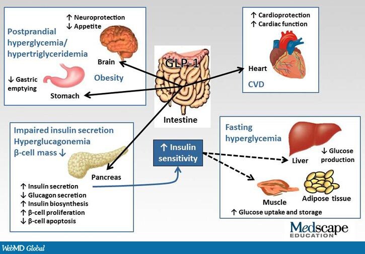
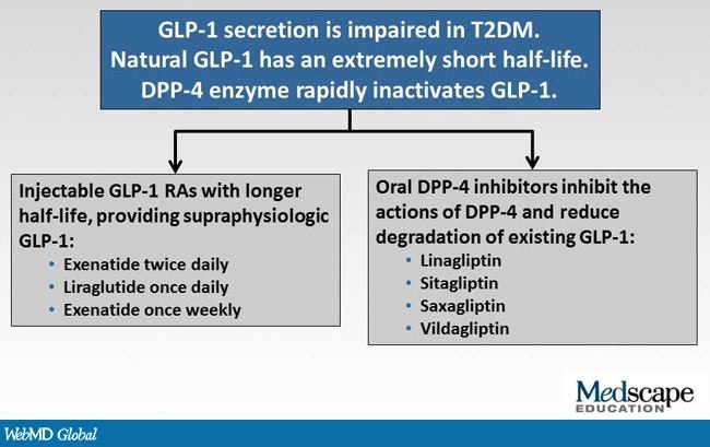
GLP-1 IN T2DM


Several antihyperglycemic agents have been developed in order to address some of the challenges associated with older agents. Incretin-based therapies emerged in the past decade and have become established as important treatment options for patients with T2DM. Two classes of agents are available: (1) DPP-4 inhibitors, which are administered orally and include sitagliptin, saxagliptin, vildagliptin, and linagliptin; and (2) GLP-1 RAs, which are administered subcutaneously and include exenatide twice-daily, exenatide once-weekly, and liraglutide.

Sodium glucose cotransporter 1 and 2 inhibitors are another class of novel antihyperglycemic agents. They inhibit glucose reabsorption from renal tubules and cause glucosuria, and sodium glucose cotransporter 1 inhibitors also lead to reduced uptake of glucose from the small intestine. Numerous sodium glucose cotransporter 2 inhibitors are undergoing preclinical and clinical trials, with dapagliflozin having completed phase 3 trials. It has been shown to reduce A1c, reduce blood glucose levels, and is associated with weight loss.


Saturday, February 2, 2013

RA TREATMENT IN PREGNANCY

Methotrexate should be avoided in women trying to conceive or who are pregnant since it is a proven teratogen (pregnancy category X). The data on biologics during pregnancy is much less clear. The anti-TNF therapies are all pregnancy category B. The other commonly used biologics in RA (abatacept, rituximab, tocilizumab) are pregnancy category C. The Food and Drug Administration (FDA) currently warns against using biologics in pregnancy. For women, who require therapy during pregnancy, glucocorticoids, hydroxychloroquine, and occasionally azathioprine (after the first trimester) are the preferred therapies for most physicians who have experience prescribing these agents during pregnancy.


Methotrexate is to be avoided, in particular, since it is a proven teratogen (pregnancy category X). It should be stopped for at least 1 menstrual cycle and as much as 6 months prior to attempting to conceive. Men with RA on methotrexate should also discontinue methotrexate for 3 months or more prior to conception. The data on biologics during pregnancy are much less clear. There have been case series describing various congenital anomalies affecting fetuses of women using these agents, and there have also been anecdotal reports of women experiencing normal pregnancies and good fetal outcomes with exposure to these compounds.[20] The anti-TNF therapies are all pregnancy category B (animal reproduction studies have failed to demonstrate a risk to the fetus, and there are no adequate and well-controlled studies in pregnant women; or animal studies have shown an adverse effect, but adequate and well-controlled studies in pregnant women have failed to demonstrate a risk to the fetus in any trimester). The other commonly used biologics for RA (abatacept, rituximab, tocilizumab) are pregnancy category C (animal reproduction studies have shown an adverse effect on the fetus and there are no adequate and well-controlled studies in humans, but potential benefits may warrant use of the drug in pregnant women despite potential risks).